

The Pitt TV Series Medical Review: Left Middle Cerebral Artery Stroke (S1E10 Review)
- Mar 18
- 8 min read

Medical dramas continually captivate audiences by exploring the absolute extremes of human physiological failure, but the most intense episodes are those that strip away the predictability of the emergency room. In its breathtaking tenth episode, The Pitt orchestrates a relentless triad of high-stakes, time-sensitive medical crises. Shifting from the delicate, microscopic neural pathways of the brain to the pressurized confines of the human eye and the catastrophic destruction of the body’s largest organ, this episode is a masterclass in extreme emergency resuscitation. Without revealing any major character arcs or plot spoilers, this comprehensive clinical review will dissect the episode’s three central, heart-pounding emergencies, the chaotic barrage of differential diagnoses, and the terrifyingly precise interventions required to pull patients back from the brink.

The Initial Presentations and the Emergency Room Visits
The clinical narrative of this episode is driven by three distinct patients, each arriving at the emergency department with presentations that require entirely different, yet equally urgent, modes of resuscitation.
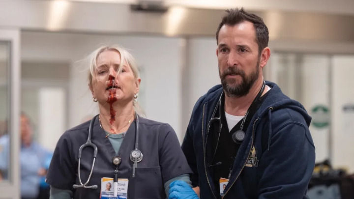
The primary medical mystery centers around Vera Mullahy, an initially unidentified "Jane Doe" in her 20s. Vera’s presentation is classically terrifying: she arrives with right-sided hemiparesis (weakness of the entire right side of her body) and a pronounced facial droop. Furthermore, she exhibits both expressive and receptive aphasia—she cannot formulate words to speak, nor can she understand what the doctors are saying to her. In the ER, these are the unmistakable, flashing red lights of a massive stroke, but her young age makes the presentation an immediate, chilling anomaly.
Contrasting Vera’s neurological silence is the visceral, localized trauma of 16-year-old Everett Young. A promising baseball pitching prospect, Everett arrives screaming in agony after taking a 100 mph line drive directly to his left eye. His presentation is a nightmare for any ophthalmologist: he has only light perception vision remaining, and his eye is visibly bulging. A rapid bedside evaluation measures his intraocular pressure at a staggering 58 mmHg (normal is between 10 and 20 mmHg).
Meanwhile, the trauma bay receives Teddy, a 28-year-old male who was caught at the epicenter of a gas tank explosion. Teddy’s presentation is a catastrophic, systemic trauma. He suffers from 90% total body surface area (BSA) burns, with the vast majority being full-thickness (third-degree) burns. His skin is charred and leather-like, and he arrives with singed nasal hairs and thick soot coating his oropharynx—a presentation that threatens to swell his airway shut at any given second.

A History Written in Impact and Lifestyle
Gathering an accurate medical history is the fundamental first step in emergency medicine, and in this episode, the histories—or the subtle clues surrounding them—are critical.
For Vera, the medical team quickly discovers she is a professional gamer. While extreme, prolonged sitting in gaming can lead to deep vein thromboses (blood clots in the legs), the history of a stroke in a 20-something typically points away from chronic issues like high cholesterol and toward structural or genetic anomalies. The historical clue here isn't necessarily her gaming, but the potential for sudden, unrecognized mechanical trauma to the neck that often plagues young stroke victims.
Everett’s history is a straightforward, high-velocity blunt force trauma. A baseball traveling at 100 mph delivers a massive kinetic payload directly to the fragile, bony orbit of the skull, virtually guaranteeing significant internal bleeding and structural fractures.
Teddy’s history is one of immediate, inescapable thermal destruction. A gas explosion not only causes severe cutaneous burns but also involves the inhalation of superheated, toxic gases, which instantly scorches the delicate mucosal lining of the respiratory tract.

Navigating the Chaos: Differential Diagnoses
The emergency department in The Pitt operates at a breakneck pace, perfectly illustrating the relentless cognitive load placed on attending physicians. They must solve the life-threatening crises of Vera, Everett, and Teddy while filtering out a constant barrage of other painful, yet lower-acuity, traumas.
While the trauma bays manage the major cases, the orthopedic and minor trauma rooms are overflowing. The doctors evaluate a non-displaced nasal fracture; though the bleeding and headache are severe, a CT scan thankfully rules out intracranial hemorrhaging and skull fractures, confirming the facial bones are intact. They manage a grueling trimalleolar ankle fracture—a highly unstable injury involving three separate bone breaks in the distal tibia and fibula. The team meticulously applies a double splint (posterior leg and sugar tong) to stabilize the mortise before prepping the patient for surgical plates and screws.
The ER board is also dotted with routine, yet time-consuming, evaluations. A patient with a split lip is assessed and safely discharged with strict instructions to avoid activities that increase cranial blood pressure. Another patient requires wound cleaning and evaluation for superficial road rash (friction abrasions) across their skin. An elbow fracture is diagnosed via X-ray, treated with standard orthopedic immobilization, and cleared for discharge. It is against this relentless background noise of broken bones and lacerations that the doctors must focus on saving Vera's brain, Everett's eye, and Teddy's lungs.

The Definitive Diagnoses: LMCA Stroke, Orbital Compartment Syndrome, and Restrictive Chest Wall
Cutting through the chaos, the medical team utilizes advanced imaging and sharp clinical acumen to arrive at the definitive, catastrophic diagnoses for their three primary patients.
For Vera, a stat CT scan of her head and neck reveals the terrifying truth. She has suffered a Left Middle Cerebral Artery (LMCA) ischemic stroke. However, the root cause is the most shocking part: she has a carotid artery dissection in the left side of her neck. The inner lining of her carotid artery tore, creating a flap where blood pooled and formed a clot. That clot subsequently broke off (embolized) and traveled directly into her left middle cerebral artery, starving the left hemisphere of her brain—which controls the right side of the body and the speech centers—of oxygen.
For Everett, the 100 mph impact caused a Grade 4 Hyphema, meaning blood has completely filled the anterior (front) chamber of his eye. Even worse, the trauma caused bleeding behind the eyeball itself. With nowhere to drain, the trapped blood pushed the eye outward, stretching the optic nerve to its breaking point and resulting in Orbital Compartment Syndrome. A subsequent CT scan also reveals a hairline orbital floor fracture.
Teddy’s diagnosis transitions from a straightforward burn case to a mechanical respiratory crisis. Because of the singed nasal hairs and soot, the doctors correctly preemptively intubate him to protect his airway from swelling shut. However, later in the episode, Teddy experiences sudden respiratory distress. His oxygen drops, peak airway pressures skyrocket, and his tidal volume plummets. The definitive diagnosis is a restrictive chest wall. His 90% full-thickness burns have destroyed the elasticity of his skin. The charred tissue (eschar) acts like a rigid, leather straightjacket, physically preventing his chest wall from expanding and crushing his lungs.
Etymology of the Diagnoses
The medical terminology in this episode is vividly descriptive. "Aphasia" comes from the Greek a- (without) and phasis (speech). "Hyphema" originates from the Greek hypo (under) and haima (blood), referring to blood settling in the eye. "Escharotomy" combines the Greek eskhar (scab or hearth) with -tome (incision), accurately describing the surgical cutting of burnt, scab-like tissue.
Understanding the Pathophysiology
The pathophysiology of Vera's LMCA stroke hinges on the concept of the "ischemic penumbra." When the clot blocked her artery, the core brain tissue died quickly, but a larger surrounding area of tissue (the penumbra) remained stunned and barely alive, desperately waiting for blood flow to be restored. Her expressive and receptive aphasia occurred specifically because the LMCA supplies Broca's area (speech production) and Wernicke's area (speech comprehension).
In Everett’s orbital compartment syndrome, the pathophysiology is a crisis of volume and pressure. The bony orbit of the skull is a fixed space. When blood rapidly fills this space, the pressure skyrockets. This immense pressure compresses the central retinal artery, starving the retina of blood, and physically stretches the optic nerve. If the pressure isn't released within 90 to 120 minutes, permanent blindness is guaranteed.
In Teddy’s case, third-degree (full-thickness) burns destroy both the epidermis and the dermis, leaving behind dead, inelastic tissue called eschar. When this leathery eschar encircles the torso, the mechanical force of the ventilator pushing air into the lungs cannot overcome the rigid resistance of the burnt skin. The lungs simply have no physical room to inflate.
The Epidemiology of the Crises
While strokes are predominantly a disease of the elderly, carotid artery dissection is one of the leading causes of stroke in young adults (under 50), accounting for up to 20% of cases in this demographic. Grade 4 hyphemas and orbital compartment syndromes are rare but well-documented emergencies, most frequently seen in high-velocity sports injuries or severe facial assaults. Severe burns covering >80% BSA carry a profoundly grim prognosis; despite advanced burn care, mortality remains exceptionally high, primarily due to the inevitable onset of systemic sepsis and multi-organ failure.

The Life-Saving Treatments Administered

The interventions showcased in this episode highlight the extreme, specialized procedures required to reverse catastrophic physiological failures.
For Vera, because she arrived within the critical four-hour treatment window, doctors administer the clot-busting drug Tenecteplase (TNK). TNK is a tissue plasminogen activator that chemically dissolves the clot in her brain. However, Vera suffers a rare, life-threatening allergic reaction to the TNK, developing angioedema (severe swelling of the lips and tongue, accompanied by a high-pitched breathing sound called stridor). The team brilliantly manages this airway emergency using a targeted pharmacological cocktail of IV epinephrine, diphenhydramine (Benadryl), and Solumedrol (a steroid) to reverse the anaphylaxis. The clot-buster ultimately works, restoring her speech and motor functions.
Everett’s treatment requires a visceral, bedside surgical procedure. To save his vision, the doctors perform a lateral canthotomy and cantholysis. Using scissors, they make a small incision at the outer corner of his eyelid (the lateral canthus) and snip the underlying tendon. This instantly allows the eyelid to hinge forward, releasing the immense, trapped pressure from behind the eye. His pressure drops, and he is rushed to ophthalmology for definitive drainage.
Teddy’s crashing respiratory status dictates a brutal but necessary intervention. To allow his lungs to inflate, the doctors perform an emergency escharotomy. Without needing anesthesia (because full-thickness burns destroy all nerve endings), they use a scalpel to make deep vertical and horizontal grid incisions directly through the charred skin of his chest. This physically releases the "straightjacket" tension, immediately allowing his chest wall to expand and restoring his ventilation, though his overall prognosis remains tragic.

A Curious Medical Fact: The "Beauty Parlor Stroke"
A fascinating clinical phenomenon related to Vera's carotid artery dissection is the concept of the "Beauty Parlor Stroke." In young, otherwise healthy individuals, the inner lining of the carotid or vertebral arteries can tear due to seemingly innocuous mechanical trauma or hyperextension of the neck. There are heavily documented cases in medical literature of individuals suffering strokes after having their necks extended backward over a salon sink for prolonged hair washing, riding intense roller coasters, practicing aggressive chiropractic manipulation, or even performing extreme yoga poses. This minor mechanical stretching creates the tear (dissection) that eventually forms the clot, proving that strokes do not solely rely on decades of poor diet and high blood pressure.

🔖 Key Takeaways
🗝️ Carotid artery dissection is a leading cause of ischemic strokes in young adults, often triggered by minor mechanical trauma to the neck that creates a blood clot which travels to the brain.
🗝️ The Left Middle Cerebral Artery (LMCA) supplies the brain's language centers; a stroke here typically causes right-sided weakness and severe expressive/receptive aphasia.
🗝️ Tenecteplase (TNK) is a powerful, time-sensitive clot-busting drug for strokes, but it carries rare risks of severe allergic reactions like angioedema, which requires immediate reversal with epinephrine.
🗝️ Orbital Compartment Syndrome is a blinding ocular emergency where trapped blood pushes the eye outward; it is treated with a bedside lateral canthotomy to snip the eyelid tendon and release the pressure.
🗝️ Full-thickness (3rd-degree) burns destroy skin elasticity, creating a rigid "eschar." If this encircles the chest, it prevents breathing.
🗝️ An emergency escharotomy involves making deep incisions through burnt tissue to physically release tension and allow the lungs to expand mechanically.
Keywords: The Pitt Medical Review S1E10
The Pitt Medical Review S1E10


Comments