

The Resident TV Series Medical Review: Subdural Hematoma, Early-Stage Gallbladder Cancer (S1E04 Review)
- Apr 6
- 8 min read

Medical television dramas often rely on massive, multi-car pileups or dramatic surgical anomalies to generate tension. However, the most chilling episodes are frequently those that highlight the quiet, insidious nature of human error and the terrifying speed at which a seemingly stable patient can decompensate. In any hospital, the emergency room triage desk is the frontline of defense; it is the critical juncture where split-second decisions dictate who lives and who dies. The fourth episode of this medical drama series masterfully exposes the vulnerabilities of this system, specifically when a hospital is severely understaffed. Without revealing the broader narrative arcs of the season, this review will examine the heartbreaking consequences of a missed neurological diagnosis and the intense surgical complexities of an unexpected oncological discovery, providing a comprehensive clinical breakdown of the episode's central cases.

Initial Presentation and the Emergency Room Visit
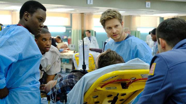
The chaos of Chastain Park Memorial Hospital's emergency department is on full display in this episode, perfectly illustrating how the sheer volume of patients can obscure critical warning signs. We are introduced to a young, initially unidentified patient named Erik. Erik arrives at the ER following a skateboard accident. Crucially, he is conscious, ambulatory, and his primary complaint is a simple headache. To an overwhelmed, inexperienced triage nurse, Erik's presentation lacks the dramatic urgency of an active hemorrhage or respiratory failure. Consequently, he is tragically categorized as a "basic" or low-priority case and sent to the waiting room.
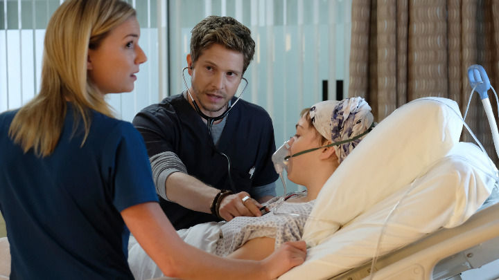
In a starkly different presentation, we meet Shirley Harris, an older woman brought into the hospital suffering from persistent abdominal pain. Unlike Erik's acute trauma, Shirley's presentation is medical in nature and initially points toward a highly common, often routine gastrointestinal complaint. The ER physician rapidly evaluates her symptoms and initially diagnoses her with simple gallstones. Both of these initial presentations represent the daily bread and butter of emergency medicine, yet both harbor deadly secrets that highlight the absolute necessity of rigorous, secondary clinical investigations.

History of Present Illness and Symptoms
The gathering of a patient's history is meant to contextualize their symptoms, but when a history is incomplete or ignored, the results are catastrophic. Erik’s history is brief but alarming to any seasoned trauma provider: he suffered a high-velocity fall from a skateboard onto a hard surface without the protection of a helmet. While his only immediate symptom is a headache, the mechanism of injury—blunt force trauma to the unprotected skull—is a glaring red flag that demands immediate advanced imaging, regardless of his initial level of consciousness.
Shirley’s history paints a more complex, chronic picture. She is an older patient presenting with localized abdominal pain and a known history of anemia (a condition where the blood lacks enough healthy red blood cells). While her abdominal pain perfectly mimics the presentation of a gallbladder attack, her underlying anemia acts as a clinical whisper, suggesting that something more systemic and insidious might be draining her physiological reserves.

The Vast Landscape of Differential Diagnoses
An emergency room is a chaotic ecosystem of competing pathologies, and the medical team must constantly sort through a vast landscape of differential diagnoses. The understaffed ER in this episode is flooded with patients requiring immediate, high-level triage.
For trauma presentations like Erik's, physicians must evaluate the severity of the impact. A patient might present with severe Road Rash (abrasions from the road surface requiring meticulous cleaning) or a Dislocated Shoulder (where the upper arm bone pops out of the socket and must be reduced). However, high-impact traumas require doctors to aggressively hunt for hidden, life-threatening injuries. They must rule out a Pneumothorax (a collapsed lung caused by air leaking into the chest cavity), Bilateral Femur Fractures (breaks in both thigh bones that carry a massive risk of fatal blood loss), or a Crushed Larynx (a severe airway obstruction potentially requiring an emergency tracheotomy). Internal organ damage is also heavily suspected; the team must look for Intra-abdominal Bleeding or a Liver Laceration. If a trauma patient exhibits muffled heart sounds and hypotension, they must act fast to treat Cardiac Tamponade, where fluid compresses the heart. For head injuries, a simple Concussion is the best-case scenario, but intracranial bleeds are the silent killers they must anticipate.
Simultaneously, the ER must manage complex medical and systemic presentations. They evaluate patients with an Essential Tremor (rhythmic muscle contractions) or Tachycardia (an abnormally rapid heart rate indicating acute stress or infection). A patient might present with Atrial Fibrillation with Rapid Ventricular Response (RVR), requiring immediate medication to control an irregular, dangerously fast heart rhythm. Less critical issues like Ear Pain or Superficial Phlebitis (vein inflammation near the skin) can clutter the waiting room, while severe threats like a Multiresistant Infection, a Rare Waterborne Sickness, or life-threatening Anaphylaxis (a severe allergic drug reaction causing airway obstruction) demand immediate pharmacological intervention. The team must also monitor metabolic derangements; for instance, Hypocalcemia (low blood calcium) can trigger a sudden Seizure. For patients like Shirley, while Gallstones are the most common cause of right upper quadrant abdominal pain, physicians must maintain a high index of suspicion to ensure these hardened deposits are not masking a far more lethal pathology.

The Definitive Diagnoses: Clinical Clues and Confirmations
The episode delivers two definitive diagnoses that hinge entirely on the passage of time and the precision of imaging technology.
For Erik, the delay in care proves fatal. Left unattended in the waiting room without a proper ID or a CT scan, his condition rapidly deteriorates from a "basic" headache into a "big deal emergency." He becomes completely unresponsive, and doctors identify a blown left pupil (a dramatically dilated pupil that does not react to light). This is the terrifying, definitive clinical sign of a massive Subdural Hematoma. The internal bleeding has accumulated to the point where the immense pressure is actively crushing his brain stem.
For Shirley, the initial diagnosis of gallstones is overturned by thorough investigative medicine. Recognizing that her clinical picture is not entirely explained by simple gallstones, the team looks closer. While the initial ultrasound missed it, a subsequent Endoscopic Ultrasound (EUS) and a CT scan reveal the horrifying truth: a hidden mass. Shirley is definitively diagnosed with Early-Stage Gallbladder Cancer, confined to the neck of her gallbladder.
Etymology of the Diagnoses
"Subdural" refers to the anatomical location of the bleed: "sub" meaning below, and "dura" referring to the dura mater, the tough outermost membrane surrounding the brain. "Hematoma" derives from the Greek "haima" (blood) and "-oma" (denoting a swelling or tumor), literally meaning a localized swelling of blood. "Carcinoma" (cancer) originates from the Greek word "karkinos," meaning crab, chosen by ancient physicians to describe the crab-like, reaching extensions of malignant tumors into surrounding tissue.
Pathophysiology
Erik's subdural hematoma was caused by the shearing and tearing of the bridging veins located between the brain and the dura mater during his fall. Venous bleeds are typically slower than arterial bleeds, which perfectly explains his "lucid interval"—the period where he was conscious and complaining only of a headache. However, as blood slowly pooled within the enclosed, rigid space of his skull, intracranial pressure rose exponentially. Eventually, this pressure forced the brain tissue downward (uncal herniation), crushing the brain stem, which controls fundamental life-sustaining functions like breathing and consciousness.
Shirley’s gallbladder cancer began as a malignant mutation in the mucosal cells lining the inner wall of her gallbladder. While many gallbladder cancers are diagnosed late because they are asymptomatic, Shirley's tumor was caught early, still confined to the neck of the organ. However, because the gallbladder rests directly against the liver, even early-stage tumors pose an immediate threat of microscopic invasion into the liver bed, complicating the physiological and surgical landscape.
Real-World Epidemiology
Traumatic brain injuries (TBIs) are a leading cause of death and disability globally, and subdural hematomas are among the most lethal of all head injuries, carrying mortality rates that can exceed 50% if surgical intervention is delayed. Gallbladder cancer, conversely, is relatively rare but highly lethal. It accounts for only about 1.2% of all cancer diagnoses but is the most common cancer of the biliary tract. Because its symptoms mimic routine gallstones, it is frequently discovered incidentally during routine cholecystectomies (gallbladder removals), and late-stage diagnoses carry extremely poor survival rates.

Aggressive Treatments and Medical Interventions
The treatments depicted in this episode contrast the devastating limits of medical timing with the awe-inspiring capabilities of modern surgical oncology.
For Erik, the treatment protocol for a massive subdural hematoma compressing the brain stem requires immediate surgical decompression, typically achieved by drilling burr holes into the skull to evacuate the pooling blood and relieve the pressure. Tragically, because he was mis-triaged and left unattended without imaging or identification, the window of opportunity to perform these life-saving burr holes closed. The prolonged compression of his brain stem resulted in irreversible damage, and Erik ultimately passed away, a victim of systemic hospital failure rather than medical impossibility.
Shirley’s treatment, however, showcases aggressive, highly skilled intervention. Due to her age, anemia, and the risk profile of the cancer, she undergoes a complex tumor resection. To ensure the cancer has not microscopically spread, the surgical team cannot merely remove the gallbladder; they must remove a portion of her liver (a liver wedge resection) to guarantee "clean margins." Furthermore, she receives experimental intraoperative radiation therapy to obliterate any remaining malignant cells in the surgical bed. The surgery becomes a high-stakes tightrope walk when the team discovers her atypical anatomy: a common hepatic artery lying completely out of its normal position. One wrong cut to this aberrant artery would have caused catastrophic bleeding. Through meticulous surgical navigation, the team successfully works around the anomaly, achieving clean margins, clear lymph nodes, and a curative outcome.

A Curious Clinical Fact: The Mechanism of a "Blown Pupil"
When the doctors identify Erik's "blown left pupil," they are witnessing a fascinating, albeit terrifying, anatomical chain reaction known as uncal herniation. The pupil's size and its ability to constrict in response to light are controlled by the oculomotor nerve (Cranial Nerve III). This vital nerve runs directly alongside the brain stem. When a subdural hematoma expands, it pushes the temporal lobe of the brain (specifically a part called the uncus) downward. This herniating brain tissue violently pinches and compresses the oculomotor nerve against the base of the skull. Because the nerve fibers that cause the pupil to constrict are located on the outside of this nerve bundle, they are the first to be crushed. The pupil instantly loses its ability to constrict, resulting in a fixed, widely dilated, or "blown" pupil on the same side as the brain bleed.

🔖 Key Takeaways
🗝️ Triage errors are fatal: Labeling a blunt-force trauma patient as a low-priority case without a CT scan can cause physicians to miss the critical window for life-saving neurosurgical intervention.
🗝️ The "Lucid Interval" is deceptive: Patients with subdural hematomas can appear perfectly conscious and coherent for hours after an injury before rapidly deteriorating as intracranial pressure reaches a tipping point.
🗝️ Burr holes relieve pressure: The primary surgical treatment for a massive intracranial bleed is drilling into the skull to evacuate the blood and prevent the brain stem from being crushed.
🗝️ Gallstones can hide cancer: Routine symptoms of gallbladder disease can easily mask early-stage gallbladder carcinoma, necessitating highly thorough diagnostic imaging like Endoscopic Ultrasounds.
🗝️ Clean margins require aggressive resection: Curing gallbladder cancer often demands removing adjacent liver tissue to ensure no microscopic malignant cells are left behind in the tumor bed.
🗝️ Anatomical variations are surgical landmines: Surgeons must be constantly prepared for atypical anatomy, such as aberrant hepatic arteries, which can turn a standard tumor resection into a life-threatening hemorrhage risk.
🗝️ A blown pupil is an extreme emergency: A fixed, dilated pupil in a trauma patient indicates active uncal herniation and impending brain death due to the compression of the oculomotor nerve and brain stem.
Keywords: The Resident Medical Review S1E04
The Resident Medical Review S1E04


Comments